|
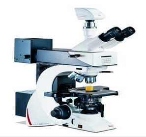
Leica Microscope |
||||
|
Optical microscopy is the oldest and most utilized method for microstructural evaluation of materials. Optical Microscopy uses the geometry and refractive indices of transparent optics to focus light and induce a magnification. The optical microscope (Leica DM2500 M) is established in the Western lab from a DST grant for the micro-fabrication lab. Basic Principle:The objective lens is, at its simplest, a very high powered magnifying glass i.e. a lens with a very short focal length. This is brought very close to the specimen being examined so that the light from the specimen comes to a focus about 160 mm inside the microscope tube. This creates an enlarged image of the subject. This image is inverted and can be seen by removing the eyepiece and placing a piece of tracing paper over the end of the tube. By carefully focusing a brightly lit specimen, a highly enlarged image can be seen. It is this real image that is viewed by the eyepiece lens that provides further enlargement. Unique Features:Fully integrated and fully automated x, y and z direction stage. Fluorescence bulb is also attached for biological applications. Upto 20X magnification. |
||||
|
Location: |
||||
|
Department of Chemical Engineering, |
||||
|
Contact: |
||||
|
Prof. Deepak Kunzru This email address is being protected from spambots. You need JavaScript enabled to view it. |
||||
|
Figure: 1 |
||||
|
Figure: 2 |
||||